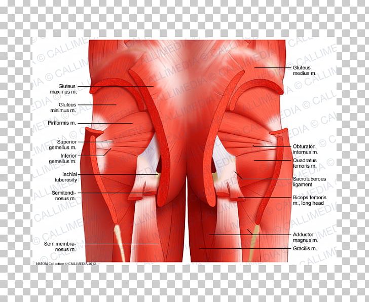
Anatomy Muscles Pelvis / The muscles that make up the pelvis diaphragm are the piriformis, coccygeus, iliococcygeus, pubococcygeus, and puborectalis.
byAdmin-
0
Anatomy Muscles Pelvis / The muscles that make up the pelvis diaphragm are the piriformis, coccygeus, iliococcygeus, pubococcygeus, and puborectalis.. The root contains three erectile tissues (two crura and bulb of the penis), and two muscles (ischiocavernosus and bulbospongiosus). To support the abdominal and pelvic viscera Use the mouse scroll wheel to move the images up and down alternatively use the tiny arrows (>>) on both side of the image to move the images.>>) on both side of the image to move the images. Continence, then pelvic muscle exercises may be effective. Muscles that attach from the pelvis to the trunk and cross the lumbosacral joint muscles that attach from the pelvis to the thigh/leg and cross the hip joint pelvic floor muscles that are located wholly within the pelvis
The ilium, ischium and the pubic bone. The levator ani muscles are the largest group of muscles in the pelvis. The pelvic girdle and pelvic spine. Below the sacrum is the coccyx, or tailbone, a section of fused bone that is the end of the vertebral. It is a broad flat muscle.
Pelvis Muscle Anatomy Muscular System Hip Png Clipart Abdomen Active Undergarment Anatomy Bone Buttocks Free Png from cdn.imgbin.com Choose from 500 different sets of flashcards about anatomy muscles pelvis on quizlet. On the other hand, if portions of those muscles are irretrievably lost, for example, due to complete. Arcus tendineus levator ani and the ischial spine The inferior aspect of the pelvic cavity is called the pelvic diaphragm. Use the mouse scroll wheel to move the images up and down alternatively use the tiny arrows (>>) on both side of the image to move the images.>>) on both side of the image to move the images. The pubic symphysis and the sacroiliac joint, and reinforced by pelvic muscles. The floor of the pelvis is made up of the muscles of the pelvis, which support its. The muscles can act separately.
Pelvic bones are held together by the two main joints of the pelvis;
The pelvic diaphragm is the third deepest layer of the pelvic floor which puts it at the very center of all the other muscles. It takes origin from the inner aspect of pelvis along a line extending from the body of the pubis to the ischial spine. These muscles origin in continuity from the body of the pubis, along a tendinous arch over the obturator internus fascia, and the ischial spine. The sacrum, five fused vertebral bones, joins the pelvis between the crests of the ilium. Über 7 millionen englischsprachige bücher. To support the abdominal and pelvic viscera The largest of them is the most superficial muscle, the gluteus maximus. In this video, we explore the anatomy of the pelvic diaphragm muscles of the pelvic floor, The floor of the pelvis is made up of the muscles of the pelvis, which support its. The muscles of the pelvic floor are collectively referred to as the levator ani and coccygeus muscles. This mri male pelvis axial cross sectional anatomy tool is absolutely free to use. Each hip bone, in turn, is firmly joined to the axial skeleton via its attachment to the sacrum of the vertebral column. The right and left hip bones also converge anteriorly to attach to each other.
The plantaris is a relatively small muscle with an appreciably long tendonous portion. Muscles that attach from the pelvis to the trunk and cross the lumbosacral joint muscles that attach from the pelvis to the thigh/leg and cross the hip joint pelvic floor muscles that are located wholly within the pelvis The sacrum, five fused vertebral bones, joins the pelvis between the crests of the ilium. The muscles of the pelvis and hip control the vast range of movement of the legs and torso. Use the mouse scroll wheel to move the images up and down alternatively use the tiny arrows (>>) on both side of the image to move the images.>>) on both side of the image to move the images.
The Muscles That Control The Pelvic Floor Pericoach from www.pericoach.com The four groups are the anterior group, the posterior group, adductor group, and finally the abductor group. Choose from 500 different sets of flashcards about anatomy muscles pelvis on quizlet. The ilium, ischium and the pubic bone. These muscles, including the gluteus maximus and the hamstrings, extend the thigh at the hip in support of the body's weight and propulsion. It helps maintain erect posture, abducts the thigh, and rotates the thigh outward. The bony framework of the pelvis is called the pelvic girdle.it is composed of the two hip bones and the sacrum. The muscles of the pelvic floor are collectively referred to as the levator ani and coccygeus muscles. The muscles can act separately.
The pelvic girdle (hip girdle) is formed by a single bone, the hip bone or coxal bone (coxal = hip), which serves as the attachment point for each lower limb.
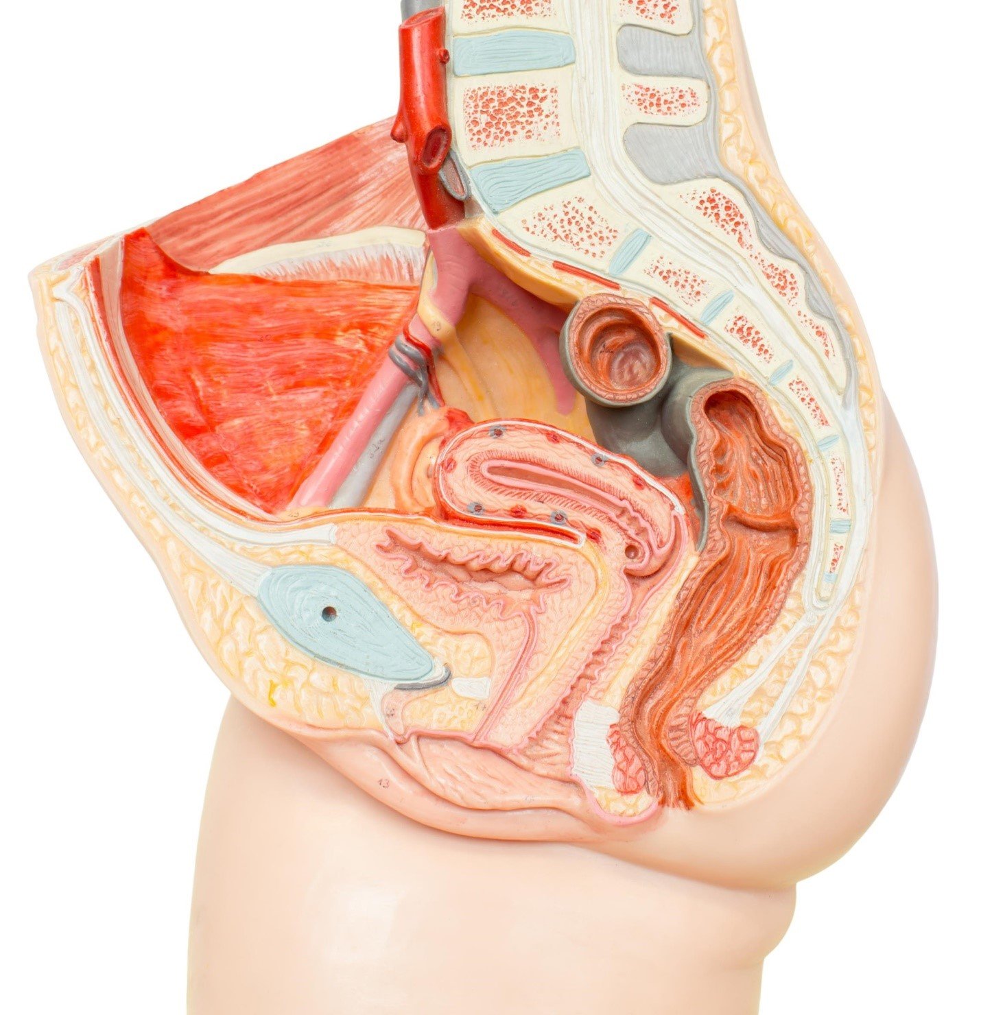
The tendinous portion can easily be mistaken for a nerve. The right and left hip bones also converge anteriorly to attach to each other. Whenever someone talks about the pelvic floor in general, they are probably talking about these 5 muscles: The pelvic cavity opens superiorly to, and is continuous with, the abdominal cavity through the pelvic inlet. The pelvic diaphragm is the third deepest layer of the pelvic floor which puts it at the very center of all the other muscles. Use the mouse scroll wheel to move the images up and down alternatively use the tiny arrows (>>) on both side of the image to move the images.>>) on both side of the image to move the images. It is located in the superficial perineal pouch of the pelvic floor, and is not visible externally. Folge deiner leidenschaft bei ebay! The pelvis is the lower portion of the trunk, located between the abdomen and the lower limbs. The levator ani muscles are the largest group of muscles in the pelvis. The pelvic girdle, also known as the hip bone, is composed of three fused bones: The pelvis also houses the reproductive organs, which have their own muscles. In this video, we explore the anatomy of the pelvic diaphragm muscles of the pelvic floor,
The bony framework of the pelvis is called the pelvic girdle.it is composed of the two hip bones and the sacrum. Below the gluteus maximus is the smaller gluteus medius. The pelvic floor muscles provide foundational support for the intestines and bladder. The pelvic girdle and pelvic spine. Folge deiner leidenschaft bei ebay!
Pelvis Muscles Of The Hip Abdomen Human Body Pelvis Hand Human Anatomy Png Pngwing from w7.pngwing.com In this video, we explore the anatomy of the pelvic diaphragm muscles of the pelvic floor, The pelvis also houses the reproductive organs, which have their own muscles. Attached to the pelvis are muscles of the buttocks, the lower back, and the thighs. The many muscles of the hip provide movement, strength, and stability to the hip joint and the bones of the hip and thigh. The levator ani muscles are the largest group of muscles in the pelvis. They have several functions, including helping to support the pelvic organs. Attached to the pelvis are muscles of the buttocks, the lower back, and the thighs. The muscles that make up the pelvis diaphragm are the piriformis, coccygeus, iliococcygeus, pubococcygeus, and puborectalis.
The muscles can act separately.
It is located in the superficial perineal pouch of the pelvic floor, and is not visible externally. The levator ani muscles consist of three. Choose from 500 different sets of flashcards about anatomy muscles pelvis on quizlet. The levator ani is a broad sheet of muscle. The plantaris muscle arises from the lateral supracondylar line of the femur and is completely absent in up to 10% of the population. The bony framework of the pelvis is called the pelvic girdle.it is composed of the two hip bones and the sacrum. Folge deiner leidenschaft bei ebay! Describe the muscles of pelvic diaphragm. The floor of the pelvis is formed by the two muscles named levator ani and coccygeus. The right and left hip bones also converge anteriorly to attach to each other. The psoas major muscle stabilizes the lumbar spine during the sitting position and the flexion of the thigh in a supine position or standing. The main function of the pelvic floor muscles are: Below the gluteus maximus is the smaller gluteus medius.